

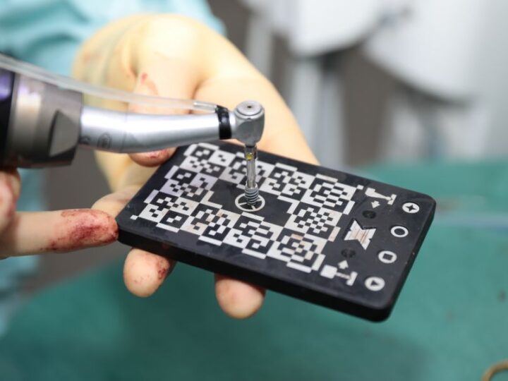
 Digital dentistry Guided vs freehand implant surgery: what is the difference?
Static guides, dynamic navigation and freehand placement compared, with the situations where each approach makes sense.
Digital dentistry Guided vs freehand implant surgery: what is the difference?
Static guides, dynamic navigation and freehand placement compared, with the situations where each approach makes sense.
Digital dentistry
What is a CBCT scan and why your surgeon needs one
What does CBCT stand for?
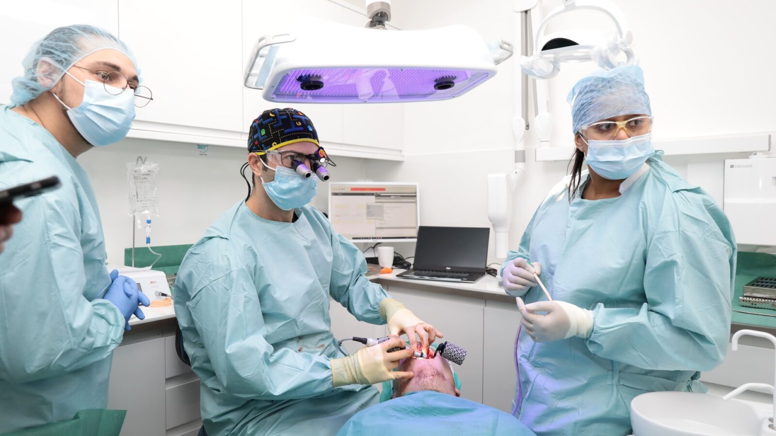
CBCT stands for cone beam computed tomography. A CBCT scan is a type of X-ray imaging that produces a three-dimensional picture of the teeth, jaws and surrounding structures. Instead of a single flat image, the scanner rotates around the head once, capturing many images from different angles. Software then reconstructs them into a 3D volume, which the clinician can view from any direction and slice at any level.
CBCT is related to medical CT scanning, but manufacturers design it specifically for the head and neck. The equipment is more compact, the scan is quicker and, for most dental purposes, the radiation dose is lower than a conventional medical CT of the same area.
How is a CBCT scan different from a normal dental X-ray?
Standard dental X-rays, such as small intraoral films or a panoramic X-ray (often called an OPG or OPT), show the teeth and jaws as a flat, two-dimensional image. Structures overlap, and measurements can be misleading. A panoramic X-ray cannot show the width of the jawbone, and it cannot reliably show whether a nerve sits in front of, behind or between the roots of a tooth.
| Intraoral or panoramic X-ray | CBCT scan | |
|---|---|---|
| Image type | Two-dimensional | Three-dimensional |
| Bone width | Not visible | Measured accurately |
| Relationship of roots to nerve or sinus | Suggested, sometimes unclear | Shown in all planes |
| Radiation dose | Low | Higher than a single X-ray, varies with field size and settings |
| Typical use | Routine checks, decay, general assessment | Implant planning, complex surgery, specific diagnostic questions |
Why does an implant surgeon need a CBCT scan?
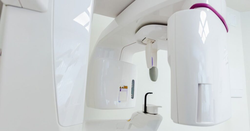
Your surgeon must place an implant within the jawbone, at a safe distance from the nerve in the lower jaw, the sinus in the upper jaw and the roots of neighbouring teeth. A CBCT scan shows exactly how much bone is available in height and width, its density and its shape. With that information, the surgeon can:
- choose the length and diameter of each implant before surgery
- identify whether you need bone grafting or a sinus lift
- locate the inferior alveolar nerve and the mental nerve precisely
- plan the angle of the implant so the final crown sits in the right position
- design a surgical guide for guided implant surgery or prepare a navigation plan
For these reasons, most implant surgeons regard a CBCT scan as an essential part of implant planning, particularly in the back of the mouth and where bone may be thin.
Which oral surgery cases need 3D imaging?
Beyond implants, surgeons use CBCT when a two-dimensional X-ray raises a question it cannot answer. Common examples include:
- lower wisdom teeth whose roots appear close to or overlapping the nerve canal on a panoramic X-ray
- impacted canines, to show their exact position and any effect on neighbouring roots
- cysts and other lesions in the jaw, to assess their size and relationship to surrounding structures
- retained roots or teeth near the sinus
- assessment before certain root canal surgeries
Your clinician always decides on clinical grounds. The team does not take a CBCT scan routinely for every patient. However, when there is a clear reason, it can make surgery safer and more predictable.
Is a CBCT scan safe, and how much radiation does it use?
CBCT uses ionising radiation, as all X-rays do. The dose varies widely depending on the size of the area scanned (the field of view), the resolution and the machine settings. A small scan of a few teeth involves much less radiation than a scan of both jaws. In general, a dental CBCT involves more radiation than a single dental X-ray but usually less than a medical CT of the head.
UK law, through the Ionising Radiation (Medical Exposure) Regulations, governs the use of X-rays. The regulations require a clinical justification for every exposure and a dose as low as reasonably practicable. In practice, the team takes a scan only when it could change the diagnosis or treatment. The field of view also covers only the area that needs assessing. The NHS guide to X-rays explains radiation doses in general terms. Let the clinic know if you are or might be pregnant.
What happens during a CBCT scan?
A CBCT scan is quick and painless. The typical process is:
- You remove metal items such as earrings, glasses, hairpins and removable dentures.
- You stand or sit in the scanner, and the team positions your head with a chin rest and sometimes a bite block.
- Then you keep still while the arm rotates around your head, which usually takes well under a minute.
- Software reconstructs the images, and they are ready to review shortly afterwards.
The scanner is open, so it is generally comfortable for people who feel uneasy in enclosed spaces.
Who reviews the images afterwards?
A trained clinician examines the whole scan and writes a report. The surgeon may need only one specific area, but the clinician still reviews the whole volume. This matters because the scan can occasionally show incidental findings, such as sinus thickening or unexpected lesions. The team explains any relevant finding to you and, where appropriate, tells your dentist or GP.
For implant planning, the surgeon usually combines the CBCT with an intraoral scan of the teeth in planning software. This allows the surgeon to design the future crown or bridge and place the implant beneath it on screen. Dr Battista uses this combined digital workflow, described on the digital dentistry page, to plan implant and surgical cases.
How does a CBCT scan support guided implant surgery?
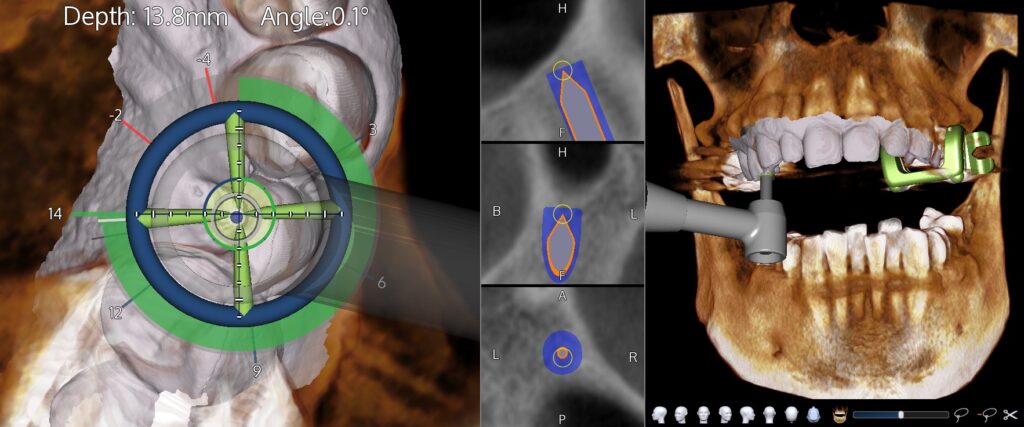
In guided surgery, the CBCT is the foundation of the whole plan. The surgeon aligns the scan with a digital impression of the teeth, designs the future tooth, positions the implant beneath it and checks the distance to every important structure. The team then turns that virtual plan into a printed surgical guide or loads it into a dynamic navigation system that tracks the drill in real time during surgery.
Without accurate three-dimensional data, neither form of guidance is possible. The quality of the scan therefore matters: movement during scanning, metal artefacts from crowns or fillings and an incorrect field of view can all reduce accuracy. A well-taken scan, checked carefully before planning, gives the surgeon a reliable map of the patient’s anatomy.
What are the limits of 3D dental imaging?
A CBCT is excellent for bone and teeth but much less useful for soft tissues. It does not show gum inflammation, the thickness of gum tissue in detail or the inside of soft tissue lesions. Metal restorations can create streaks that obscure nearby areas. For these reasons, clinicians always interpret CBCT alongside a clinical examination and, where appropriate, other investigations such as periodontal charting or a biopsy.
Can your dentist share existing images?
Yes. If you have had a recent CBCT of the relevant area, sharing the original data files, rather than screenshots, can avoid a repeat exposure. Whether you need a new scan depends on its age and on any changes since, such as an extraction or a graft. It also depends on whether the field of view covers the treatment area. Dentists referring patients can include existing images with the referral.
How should you prepare for the appointment?
You need very little preparation. Wear comfortable clothing without metal near the neck, and be ready to remove earrings, piercings in the mouth or face, glasses and any removable dentures or retainers. Mention any recent scans of the head or jaws, and bring the files if you have them. If you find it hard to keep still, let the team know so they can position you comfortably before the scan starts.
Is a CBCT scan always necessary before an implant?
For almost all implant cases, surgeons recommend three-dimensional imaging to assess bone volume and anatomy properly. A few very straightforward sites may call for a different judgement, but surgeons increasingly consider a CBCT a standard part of safe implant planning. Your surgeon will explain why you need a scan and what it should show.
If your dentist has advised a CBCT scan, or you would like an assessment for implants or oral surgery, you can contact the clinic to discuss it.
Frequently asked questions
Does a CBCT scan hurt?
No. A CBCT scan is completely painless. You stand or sit still while the scanner arm rotates around your head, which usually takes well under a minute. You may be asked to bite gently on a small block to keep your teeth in position. The scanner is open, so most people find it comfortable, including those who dislike enclosed spaces.
How much radiation does a dental CBCT involve?
The dose depends on the size of the area scanned and the machine settings. A dental CBCT generally involves more radiation than a single dental X-ray but usually less than a medical CT scan of the head. UK regulations require every scan to be clinically justified and the dose kept as low as reasonably practicable, so the field of view is limited to the area needed.
Can I have a CBCT scan if I am pregnant?
Elective X-ray imaging is usually postponed during pregnancy unless it is clinically necessary. Always tell the clinic if you are or might be pregnant. Most implant and elective oral surgery planning can be safely delayed until after pregnancy, and urgent problems are managed with the minimum imaging needed.
Why do I need a CBCT for wisdom tooth removal?
A CBCT is not needed for every wisdom tooth. It is recommended when a standard X-ray suggests that the roots are close to the nerve that supplies sensation to the lip and chin. The 3D scan shows the exact relationship, allowing the surgeon to assess the risk properly and, if necessary, choose a technique that reduces it.
Who reads and reports a CBCT scan?
A CBCT scan must be reviewed and reported by an appropriately trained clinician. The whole scanned volume is examined, not only the area of interest, so that incidental findings are not missed. Any relevant findings are explained to you and, where appropriate, shared with your dentist or GP.
More guides
All guides
Digital dentistry Guided vs freehand implant surgery: what is the difference?
Static guides, dynamic navigation and freehand placement compared, with the situations where each approach makes sense.
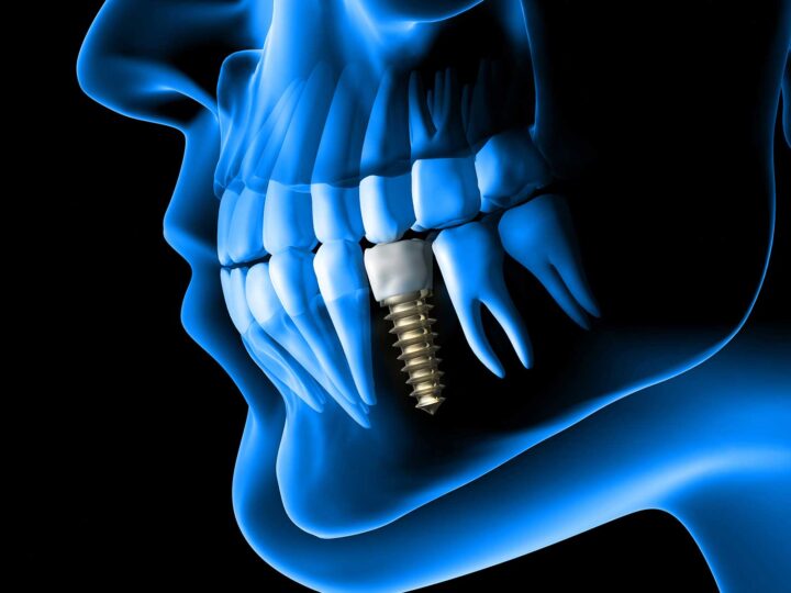 Dental implants How much do dental implants cost in London?
The real factors behind implant pricing in London, and how to compare treatment quotes on a like-for-like basis.
Dental implants How much do dental implants cost in London?
The real factors behind implant pricing in London, and how to compare treatment quotes on a like-for-like basis.
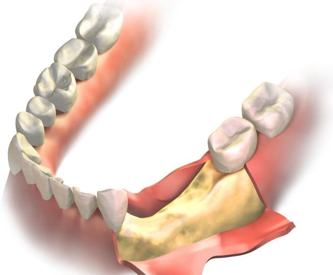 Dental implants Am I a candidate for dental implants?
How bone, gum health, medical history and smoking decide whether dental implants are suitable, and what can improve the odds.
Dental implants Am I a candidate for dental implants?
How bone, gum health, medical history and smoking decide whether dental implants are suitable, and what can improve the odds.
Next step
Talk it through before you decide
Send me a short message about what is going on. You will get an honest answer on whether surgery is the right step, and what the options are.